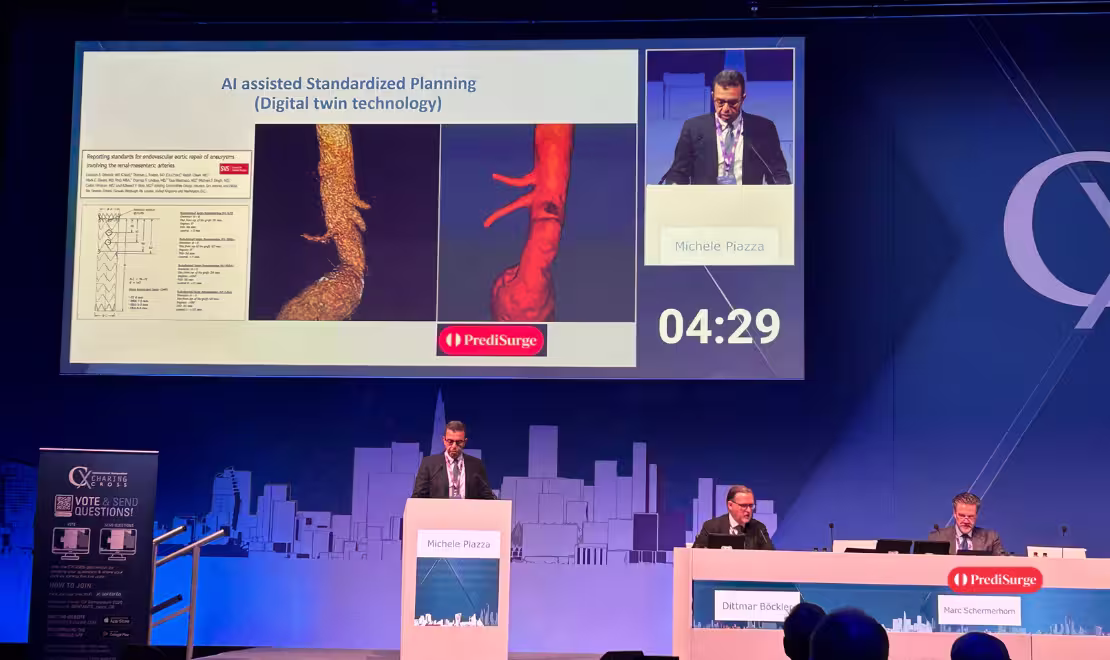
AI assisted planning for Fenestrated PMEG

Planning remains one of the most demanding steps in complex endovascular aortic repair. Unlike standard EVAR, fenestrated repair requires precise alignment between each graft fenestration and the corresponding target vessel ostium.
This involves accurately defining both the longitudinal position along the aorta and the circumferential clock position. Even minor deviations can translate into challenging cannulation, suboptimal bridging-stent angulation, or compromised long-term durability.
The problem of operator-dependent planning
Currently, planning is operator-dependent, resting on each operator’s personal experience with sizing software and CT review. No matter how skilled the planner, there is always a large number of small biases that creep into the measurements.
While experienced operators develop strong consistency, variability is never fully eliminated. Small differences in centreline orientation, ostium interpretation, or angulation correction introduce cumulative bias.
How AI actually changes planning of complex EVAR
AI assistance brings two concrete advantages:
- It limits bias and the risk of misaligning the fenestration during planning, by replacing the operator’s manual measurement choices with a consistent, reproducible measurements.
- it significantly reduces the time the operator needs to spend planning — a workflow that today can stretch across hours of CT analysis before a single complex case.
AI Powered planning — A profound shift
Prof. Piazza goes further than calling AI a mere support tool. In his view, AI-powered planning represents a profound shift in how vascular cases are approached — not by replacing the physician, but by elevating their role.
Rather than spending time on the technical work of marking and measuring on a DICOM viewer, the surgeon's contribution shifts upstream: into clinical decision-making, anatomical strategy, and case selection. AI handles the groundwork so that physicians can focus on what matters most.
Results of SPHERE Registry
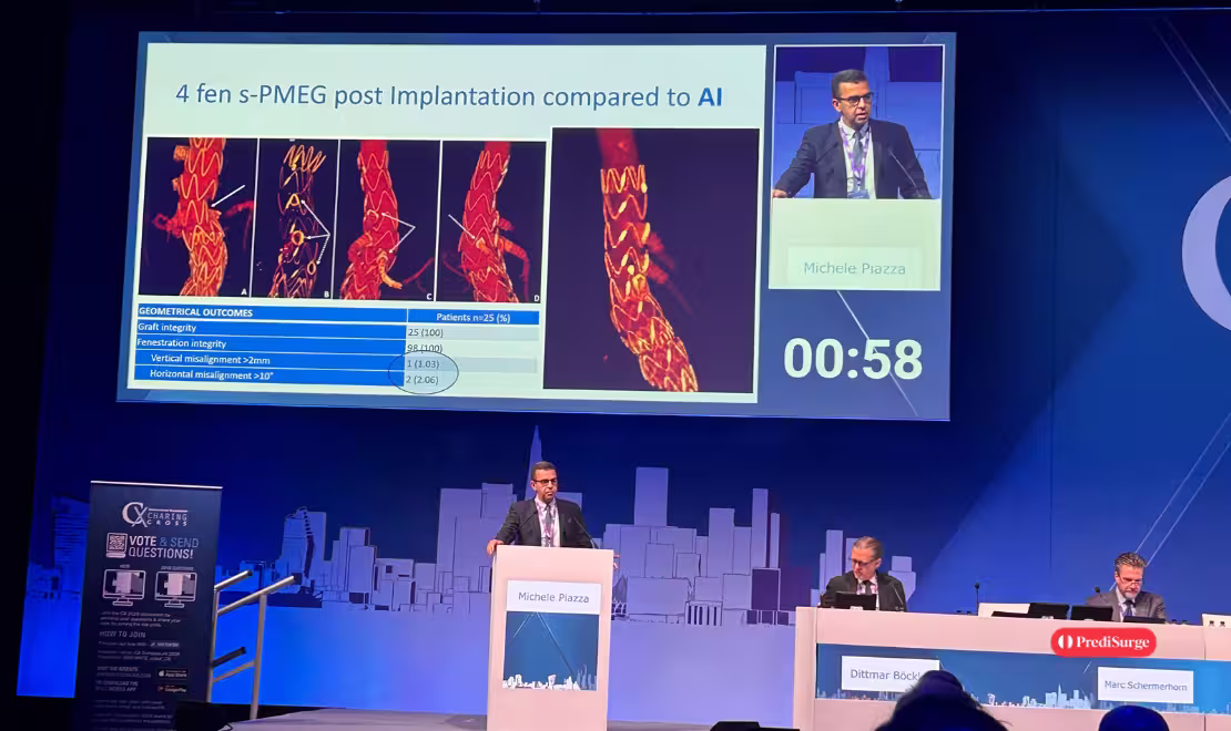
With the SPHERE registry and the standardisation of planning, The risk of misalignment at post-op is significantly reduced. In practice, this means successful access and bridging of the fenestration in all the cases — every target vessel cannulated, every bridging covered and stent deployed at a favourable angle.
Furthermore a limited long-term instability, because the fenestrations sit well in position at the ostium of the target vessel, which is the geometric precondition for durable sealing and patency of the visceral and renal branches.
Vision for PMEG using AI
The future is to put the software directly to physicians’ hands — as an app or in the browsers. The workflow would be simple: Take the pre-implantation CT, upload the images, and within about three to four minutes receive a complete report of where to place the fenestration in the graft.
For a technique that today demands one of the most time-intensive planning workflows in vascular practice, that is a meaningful shift in how complex aortic repair gets done.
Workflow reimagined with PrediSurge
The workflow collaboratively in development with PrediSurge reduces the operator’s task to two clinical decisions:
- Where they want to land the graft in the suprarenal or supra iliac aorta,
- The diameter of the graft they want to use.
From those two inputs alone, the automated software returns where to place each fenestration — both the vertical length (cranio-caudal position along the graft body) and the lateral distance (clock-face position around the circumference).
Critically for complex anatomy, the tool accounts for aortic specifics that drive deployment behaviour in the real world: angulations along the visceral segment, and diameter mismatch between the suprarenal and the pararenal aorta — a factor that is particularly relevant in patients with conical necks, asymmetric para visceral dilatation, or significant aortic tapering across the target zone.